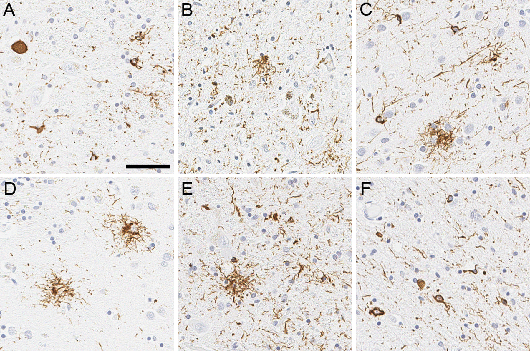
Figure 1:

Representative images of tau immunohistochemistry. Immunohistochemistry using anti-phospho-tau antibody reveals globose tangles, coiled bodies, and threads in the subthalamic nucleus (A). Neuronal loss with extraneural pigment, globose tangles, tufted astrocyte, and coiled bodies are observed in the ventrolateral cell group of substantia nigra (B). Tufted astrocytes are frequent in the motor cortex (C), caudate nucleus (D), and red nucleus (E), accompanied with coiled bodies and threads. Midbrain tectum shows numerous coiled bodies and threads (F). Scale bar = 50 μm.
