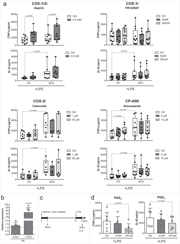
Fig. 3. Inhibition of the COX pathway does not decrease BCG-induced cytokine production.
a TNFα and IL-6 secreted by macrophages exposed to BCG and (0.5 mM) Aspirin, (30, 300 nM) FR122047, (1, 50 µM) Celecoxib or (5, 10 µM) Itraconazole for the first 24 h of culture and 5 days later stimulated with LPS for 24 h (n = 9 (Aspirin and Itraconzole), n = 12 (FR122047), n = 6 (Celecoxib) biologic replicates, pooled from 3 (Aspirin and Itraconazole), 4 (FR122047) or 2 (Celecoxib) independent experiments). b Relative COX1 and COX2 expressions in monocytes incubated with BCG for 4 h (n = 6 biologic replicates pooled from 2 independent experiments). c Schematic representation of in vitro LM supplementation experiments. d TNFα and IL-6 secreted by macrophages exposed to (10, 100 pM) PGE2 for the first 24 h of culture and 5 days later stimulated with LPS for 24 h (n = 6 biologic replicates, pooled from 2 independent experiments). a Mean, 10–90 percentile and whiskers extend to the most extreme points, b, d Mean + SD. a two-way ANOVA, Sidak’s multiple comparisons test, b two-tailed Wilcoxon matched-pairs signed rank test, d Friedman’s test followed by Dunn’s multiple comparisons test. (Ctrl control, BCG Bacillus Calmette-Guérin, LPS lipopolysaccharide, COX cyclooxygenase, CP cytochrome P, PG prostaglandin).

