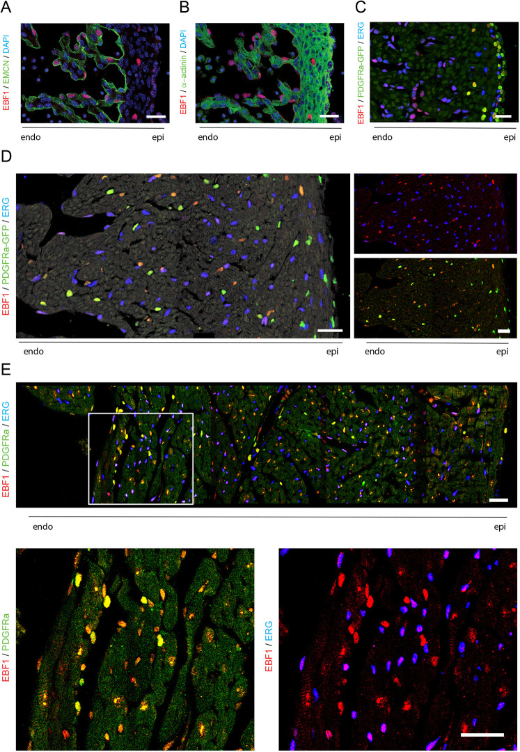
Fig. 6.
EBF1 expression in the murine heart. (A,B) Immunofluorescence of EBF1 co-stained with EMCN (A) or the myocyte marker α-actinin (B) in E13.5 embryonic heart sections. (C-E) Immunofluorescence of EBF1 co-stained with the endothelial marker ERG and the fibroblast/smooth muscle cell marker PDGFRα in heart sections at (C) E13.5, (D) postnatal day 1 and (E) postnatal day 21. High-magnification images of areas outlined in E are shown underneath. All images are at the mid level of the left ventricle. Scale bars: 25 µm.

