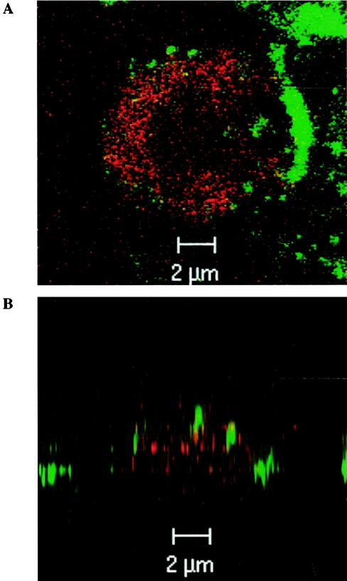
FIG. 7.
Visualization of IL-18-induced fMLP-R endocytosis by confocal microscopy. Whole-blood samples were incubated at 4°C for 1 h with FITC-anti-human CD15 and PE-anti-human fMLP-R antibodies. After the samples were washed once in ice-cold PBS, they were incubated at 37°C with IL-18 (500 ng/ml) for 45 min. Images were obtained by confocal laser scanning microscopy. (A) Horizontal (x-y) section; (B) vertical (x-z) section. Thirty optical sections were used for three-dimensional reconstruction.

