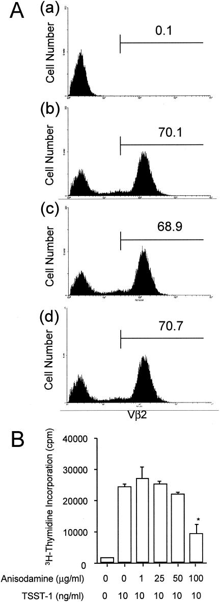
FIG. 7.
Inhibition of the TSST-1-stimulated proliferative responses of human PBMCs by anisodamine. (A) PBMCs (2 × 10⁶ cells/ml) were stimulated with TSST-1 (10 ng/ml) for 72 h in the presence or the absence of anisodamine (50 or 100 μg/ml). To allow reexpression of the T-cell receptor, the cells were further treated with rIL-2. The cells were stained with FITC-Vβ2 and PE-CD3 antibodies, and then the labeled cells were examined by flow cytometric analysis. (Part a) CD3+ cells (T cells) in PBMCs without TSST-1 treatment; (part b) CD3+ cells (T cells) in PBMCs after stimulation with TSST-1; (part c) CD3+ cells (T cells) in PBMCs after stimulation with TSST-1 in the presence of 50 μg of anisodamine per ml; (part d) CD3+ cells (T cells) in PBMCs after stimulation with TSST-1 in the presence of 100 μg of anisodamine per ml. The numbers within each panel represent the percentage of Vβ2+ T cells. (B) PBMCs were stimulated with TSST-1 (10 ng/ml) for 72 h in the presence or the absence of various concentrations of anisodamine. The cultures were then pulsed with [3H]thymidine for 16 h, and the amount of [3H]thymidine (in counts per minute) incorporated into PBMCs was measured. *, P < 0.05 compared with the levels of [3H]thymidine incorporated in the TSST-1-stimulated cells in the absence of anisodamine.

