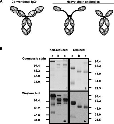
FIG. 1.
(A) Structure of camelid IgGs (data from reference 12). (B, top) Coomassie blue-stained SDS-PAGE gel of purified llama IgG isotypes resolved under nonreducing and reducing conditions. (a and b) IgG1 and IgG3 (eluted from protein G); (c) IgG2 (eluted from protein A). (Bottom) Western blot of gels identical to Coomassie blue-stained gels developed with polyclonal anti-llama IgG (H+L). Molecular masses (in kilodaltons) of protein standards run in parallel are indicated.

