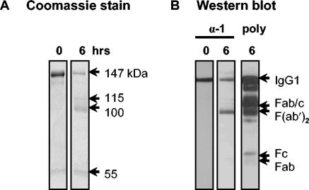
FIG. 3.
Binding of anti-llama IgG1 MAb (27E10) to fragments of llama IgG1. (A) Coomassie blue-stained gel of llama IgG1 digested for 0 or 6 h with pepsin in acetate buffer, pH 4.5. Estimated molecular masses (in kilodaltons) are indicated. (B) Western blot showing the reactivity of 27E10 (α-1) with llama IgG1, which was digested for 0 or 6 h with pepsin and developed with HRP-goat anti-mouse antibodies. poly, HRP-goat anti-llama IgG (H+L) against pepsin-cleaved IgG1.

