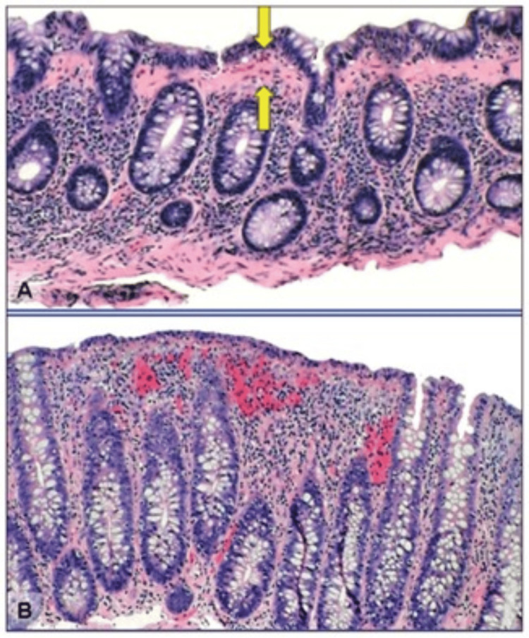
Figure 1. Histological features of microscopic colitis.
(A) Collagenous colitis is associated with an increased lymphocytic infiltrate of the lamina propria and intraepithelial lymphocytosis. There is a markedly thickened (>10 μm) collagen band underlying the colonic epithelial cells (arrows). (B) Lymphocytic colitis is associated with an increased lymphocytic infiltrate of the lamina propria and intraepithelial lymphocytosis. The collagen band underlying the colonic epithelial cells is normal in width (<10 μm) [58].

