
FIGURE 1.
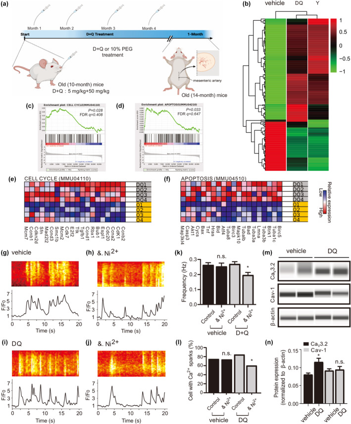
D + Q turnover caveolar Ca2+ sparks in aged mesenteric VSMCs. (a), experimental design of senolytics administration is shown. (D, dasatinib; Q, quercetin; PEG, polyethylene glycol). (b), heat map of RNA‐seq data of mesenteric artery from young, vehicle‐, and D + Q‐treatment mice (n = 4 samples for each group). (c–f) Shown are gene set enrichment analyses (c, d) and heat maps for the top 20 up‐regulated genes for CELL CYCLE (e) and APOPTOSIS (f) in D + Q compared with vehicle group. FDR, false discovery rate. (g), Ca2+ fluorescence line scan images of a Fluo‐4‐AM–loaded VSMC from a middle‐aged mouse and the time course of Ca2+ fluorescence changes. (h), same as (g) but in the presence of methyl‐β‐cyclodextrin (dextrin, 10 mM, 90 min at room temperature). (i), same as (g) but in the cell from a D + Q‐treated aged mouse. (j), same as (i) but in the presence of methyl‐β‐cyclodextrin. (k–l), summary of the results. Ca2+ spark frequency (e) and fraction of cells producing Ca2+ sparks (f) in VSMCs from aged mice (n = 134), in VSMCs from aged mice incubated with methyl‐β‐cyclodextrin (n = 95), in VSMCs from D + Q treated aged mice (n = 175), and in VSMCs from D + Q treated aged mice incubated with methyl‐β‐cyclodextrin (n = 175). Cells were isolated from 4 mice in each group. VSMC, vascular smooth muscle cell. *p < 0.05; n.s., not significant. (m), western blot analysis of CaV3.2, Caveolin‐1 proteins in mesenteric arteries of aged versus D + Q mice. (n), quantification of western blot results. Western blot results were analyzed from 8 mice in each group. *p < 0.05; n.s., not significant; Cav‐1, Caveolin‐1.