
FIGURE 1.
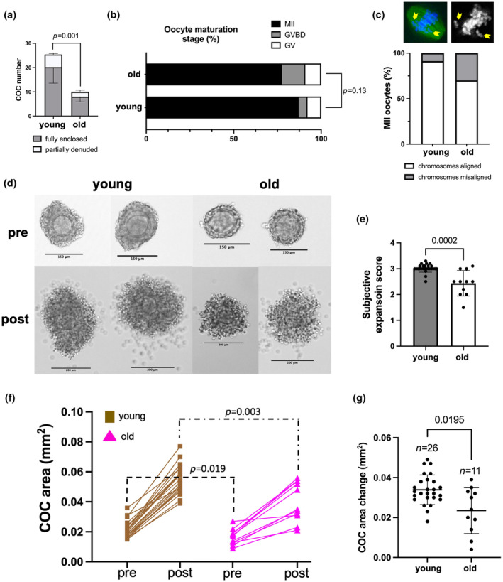
Cumulus expansion is impaired in COCs from reproductively old mice during IVM. (a) Significantly fewer COCs were obtained from reproductively old (14–17 months) mice after hormonal stimulation with PMSG. (b) Oocyte maturation stages were not significantly different between reproductively young and old mice after IVM of COCs in a conventional incubator. (c) Chromosome misalignment on metaphase II spindles showed a trend toward increased abnormalities with age (p = 0.15). (d) Representative images of COCs from reproductively young and old mice before and after cumulus expansion (scale bars: 150 μm for pre‐ and 200 μm for post‐expansion images) (e) COCs from reproductively old mice exhibited decreased subjective expansion scores. (f) COC area was decreased pre‐ and post‐expansion in COCs from reproductively old mice. (g) The change in COC area (post‐expansion area – pre‐expansion area) was significantly less in COCs from reproductively old mice during IVM (testing the difference between differences). Data are represented as mean ± SD. Experiments were repeated 5 times with the comparison of all COCs from one young and one old mouse per experiment (5–29 COCs per mouse). (b, c) Represent pooled analyses of oocytes from all experiments. Two‐sided Student's t‐test or Mann–Whitney U test was used to compare continuous variables depending on normality. Chi‐square test was used to compare categorical variables. p < 0.05 was considered statistically significant. GV, germinal vesicle; GVBD, germinal vesicle breakdown; IVM, in vitro maturation; MII, metaphase of meiosis II; PMSG, pregnant mare serum gonadotropin.