FIGURE 5.

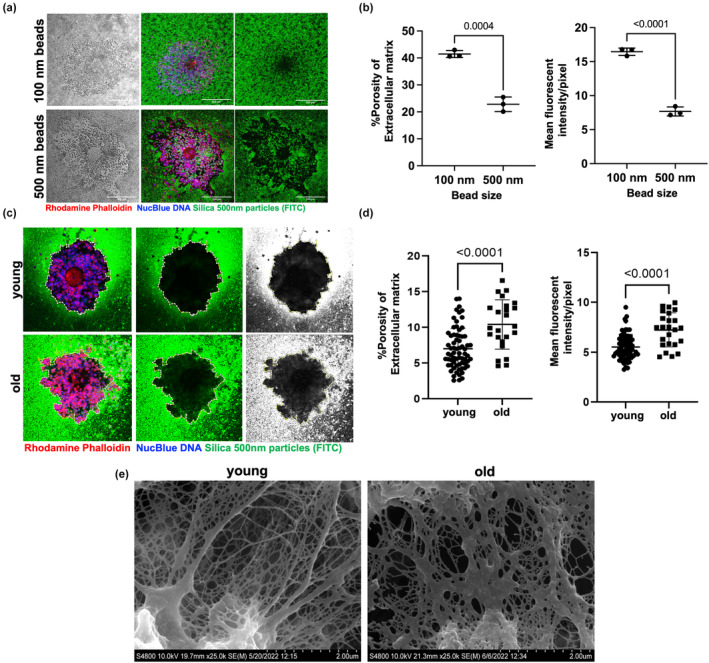
Extracellular matrix (ECM) integrity is compromised with age in expanded COCs. (a) Particle exclusion assay (PEA) was performed on expanded COCs using fluorescent silica nanoparticles of different sizes: Brightfield image (left panel); merge channel (middle panel); green channel (right panel). COCs incubated with 500‐nm nanoparticles demonstrated a clear zone of exclusion at the periphery (scale bars—200 μm). (b) ECM percent porosity (calculated by dividing the area infiltrated by nanoparticles within the COC to the whole area occupied by the COC) and nanoparticle mean fluorescent intensity/pixel of COC image (calculated as the total nanoparticle fluorescent intensity per COC/total number of pixels per COC image) provide quantitative assessment of ECM permeability to these nanoparticles. (c) Representative images of the PEA performed on COCs from reproductively young and old mice using 500‐nm nanoparticles: Merge channel (left panel); green channel (middle panel); grayscale image of the green fluorescent nanoparticles (right panel). The contrast was adjusted similarly across images to highlight particle distribution. (d) Porosity of ECM and nanoparticle mean fluorescent intensity/pixel of COC image demonstrated increased permeability to 500‐nm nanoparticles in expanded COCs of reproductively old mice (young: pooled analysis of 69 COCs from 6 mice, and old: pooled analysis of 24 COCs from 6 mice) (DNA, blue; Actin, red; silica nanoparticles, green). (e) Representative scanning electron microscopy images of expanded COCs from reproductively young and old mice highlighting the matrix region (33 COCs from 5 young and 19 COCs from 5 old mice were used, scale bars 2 μm). Data are represented as mean ± SD. Two‐sided Student's t‐test was used for statistical analysis. p < 0.05 was considered statistically significant.
