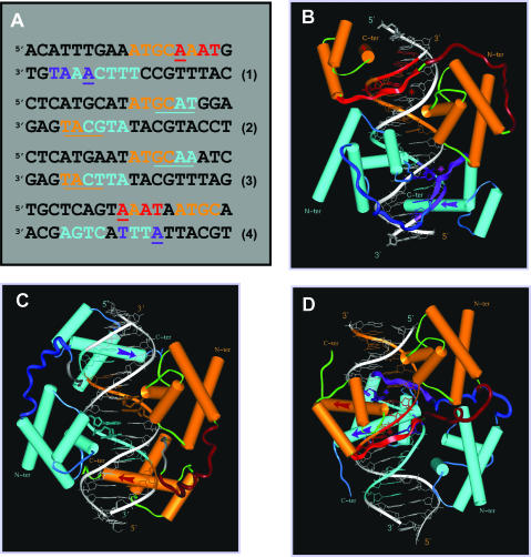
Figure 1.
The three generic modes of N-Oct-3 POU domain homodimerization. (A) Eighteen base pair DNA sequences of: (1) the osteopontin gene enhancer fragment defining the PORE motif, (2) the prototypic MORE motif, (3) the MORE-type IgG VH gene promoter canonical fragment and (4) the AADC gene promoter fragment defining the new NORE motif. In all the cases, the first and second N-Oct-3 POU domain binding sites are coded in brown and turquoise, respectively. In the PORE (1) and NORE (4) motifs, critical nucleotides of the first and second POUh tetrameric sub-sites are coded in red and indigo respectively. In the MORE motifs (2 and 3), the first and second POUh tetrameric sub-sites are underlined in brown and turquoise, respectively, to compensate for the overlap with the POUs binding sub-sites. (B–D) Modeled structures of the homodimeric complexes between the N-Oct-3 DBD and (B) a PORE motif (sequence 1), (C) a MORE motif (sequence 3) and (D) a NORE motif (sequence 4). In all the cases, the respective display-codes for the first and second N-Oct-3 POU domains are as follows: brown- and turquoise-colored cylinders for the α-helices, red- and indigo-colored arrows for the POUh recognition helices, dark brown- and blue-colored coils for the linkers. In the PORE (B) and NORE (D) types of complexes, the first and second POUh N-terminal extensions are displayed as red- and indigo-colored ribbons, respectively, their insertion into the DNA minor groove being indicated by asterisks of the identical color.

