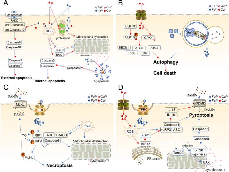
Fig. 4.
Iron and copper induce diverse types of cell death. A Iron and copper trigger external and internal apoptosis. In the internal pathway, they lead to mitochondrial dysfunction, regulate BCL−2 family proteins, and release cytochrome c through ROS and proteasome inhibition. Cytochrome c is dependent on BCL−2 family proteins that bind and activate apoptotic protease-activating factor 1 (APAF-1) as well as procaspase 9, forming an apoptosome. B Iron and copper lead to autophagy. Ferritin is degraded by autolysosomes, leading to abnormal iron accumulation and eventually triggering cell death. Copper binds to ULK1 and ULK2 directly, relieving ULK1/ULK2 inhibition and promoting autophagy. Copper also binds to GPX1 to induce autophagy. C Iron overload accelerates ROS accumulation and the RIPK1/RIPK3/MLKL pathway, opens the mitochondrial permeability transition pore (mPTP), eliminates mitochondrial membrane potential, and releases cytochrome c outside mitochondria and DAMPs to the extracellular space, resulting in definitive necroptosis. D Iron stimulates the production of ROS, which induces pyroptosis via the classical caspase-1-mediated pyroptotic pathway and caspase-3-dependent pathway. In the first pathway, iron-elevated ROS cause the oxidation and oligomerization of Tom20, which recruits Bax and facilitates cytochrome c release into the cytosol. Then, cytochrome c activates caspase-9, which activates caspase-3. Finally, caspase-3 aggravates GSDME cleavage and triggers pyroptosis. In the second pathway, iron accelerates ROS accumulation, which activates the NLRP3/ASC/Caspase1 complex and hence induces pyroptosis. Copper also stimulates the production of ROS, induces ER stress and triggers the classical Caspase-1-mediated pyroptotic pathway through the IRE1α-XBP1 axis. The red arrow indicates Cu, and the blue arrow indicates Fe

