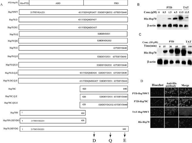
Fig 1.
Protein transduction domain (PTD)–Hsp70, TAT-Hsp70 proteins, and their mutants. (A) Diagram of Hsp70 and its mutant proteins used in this study. The wild-type Hsp70 protein (Hsp70WT) had a 6.His domain followed by the protein transduction domain (PTD or TAT) fused to the N-terminal of the human Hsp70cDNA. (B,C) Concentration- (B) and time (C)-dependence of the cellular import of PTD-Hsp70WT and TAT-Hsp70WT proteins. NT cells were treated with PTD-Hsp70WT or TAT-Hsp70WT at the concentrations indicated for 0.5 hours at 37°C (B); or the cells were exposed to 10 μM PTD-Hsp70WT or TAT-Hsp70WT proteins at 37°C for the times indicated (C). The imported proteins were immunoblotted with histidine (His) antibody. (D) Immunofluorescent staining of NT cells treated with PTD-Hsp70s or TAT-Hsp70WT. Cells were incubated for 30 minutes with 10 μM of each protein. Left panels show nuclear staining with Hoechst, the right panels show anti-His antibody staining for each of the imported proteins

