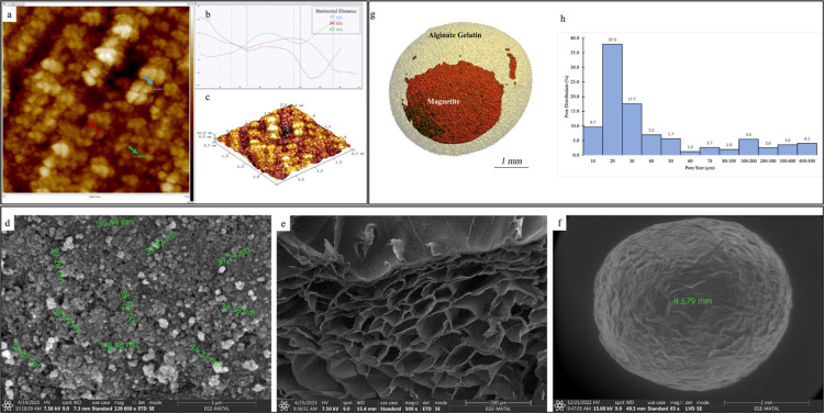
Figure 4.
(a) 2D image of AFM scan of magnetite nanoparticles, (b) measurement of diameters of nanoparticles, (c) 3D topography image of the nanoparticles, d) SEM image of magnetite nanoparticles, (e) SEM image of cross-section of the bead, (f) SEM image of the outer surface of the magnetic beads, (g) 3D model of the GA cross-linked magnetic bead created with micro-CT analysis, and (h) pore distribution histogram of beads.

