
Fig 3.
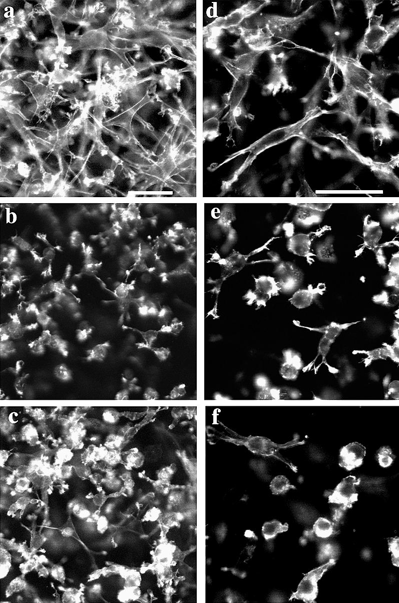
F-actin staining of cells plated in attached collagen lattice. Six hours after plating, cells were fixed and stained with BODIPY-phalloidin to visualize the actin cytoskeleton. Note that SS12 cells (a, d) were more elongated in a bipolar manner than VO cells (b, e), whereas AS10 cells (c, f) were more rounded in appearance. Bar = 50 μm