
Figure 2.
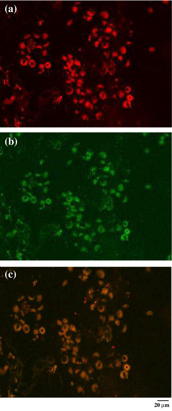
Expression of IP-10/CXCL10 in the synovium of a representative patient with JIA. Immunofluorencence confocal laser scanning microscopy indicates the presence of chemokine IP-10 (red) (a); (b) the same cells are shown to be synovial macrophages, as they are marked with CD68 (green). (c) The co-localization of IP10 and CD68 by macrophages (brown) is clearly visible. Original magnification ×1000.