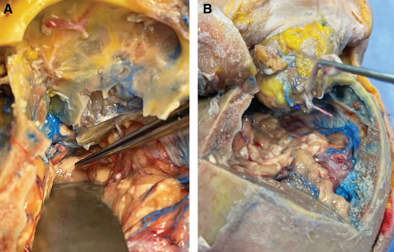
Fig. 4.
Superior view of the optic nerve after craniectomy in a cadaveric donor. Before transection, with retraction of the brain, the optic nerve (indicated by forceps) is visible immediately distal to the optic chiasm (A). After release, the optic nerve is seen alongside the ophthalmic vasculature posterior to the annulus of Zinn (B). Red silicon injection indicates arterial system and blue indicated venous. Printed with permission and copyrights retained by Eduardo D. Rodriguez, MD, DDS.

