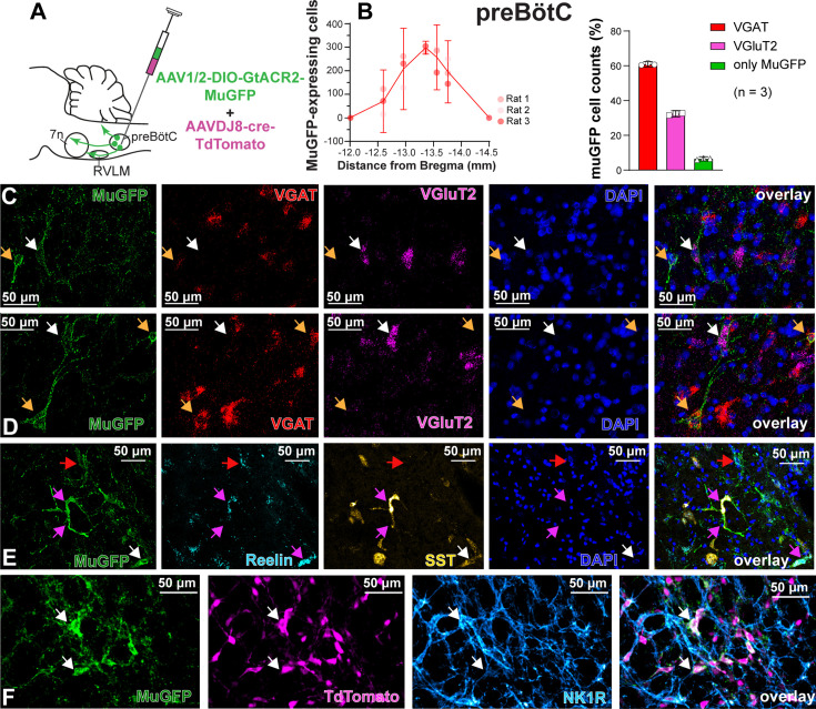
Figure 4. Excitatory and inhibitory pre-Bötzinger complex (preBötC) neurons are transduced by non-selective transfection of preBötC neurons.
(A) Schematic diagram showing injection protocol for non-selective transduction of preBötC neurons. (B) Quantification of the total number of MuGFP-expressing neurons, plotted as the distance from Bregma (mm). The histograms show the number of transduced cells that co-expressed mRNA for VGAT or VGluT2. The results are presented as mean ± 95% CI. (C–E) In situ hybridization showing the co-expression of MuGFP (green), with mRNA for VGAT (red), VGluT2 (magenta), reelin (cyan), and somatostatin (SST) (yellow) in preBötC. Nuclei are labeled in blue (DAPI). The yellow arrows highlight VGAT neurons, the white arrows highlight VGluT2 neurons, the red arrows highlight reelin neurons, and the pink arrows highlight reelin and SST neurons. (F) Immunohistochemistry showing that some neurons expressing MuGFP (green) and TdTomato (magenta) also express neurokinin-1 receptor (NK1R) (light blue). Co-localization is indicated by white arrows.

