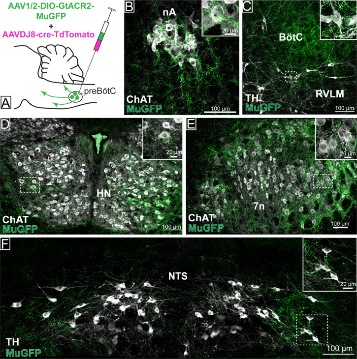
Figure 6. Distribution of GtACR2-MuGFP-expressing axons in multiple brainstem nuclei following the non-selective transduction of pre-Bötzinger complex (preBötC) neurons.
(A) Schematic diagram showing the protocol for non-selective transduction of preBötC neurons. Confocal microscopy images demonstrate MuGFP expression in axon in the (B) nucleus ambiguus. (C) Rostral ventrolateral medulla and Bötzinger complex, (D) hypoglossal nucleus, (E) facial nucleus, and (F) nucleus of the solitary tract. Higher magnification images of the hashed-boxed regions are shown in the upper right corner of the lower magnification image. ChAT: choline acetyltransferase; TH: tyrosine hydroxylase.

