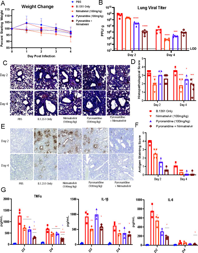
Fig 3.
Combination of nirmatrelvir and pyronaridine treatment additively reduces SARS-CoV-2 infection and inflammation in vivo. Eight- to 10-week-old wild-type BALB/c mice were treated with nirmatrelvir (oral administration), and/or nirmatrelvir (oral administration) daily at the indicated concentrations starting 12 h before infection. Mice (n = 5 per group) were intranasally inoculated with 1 × 10⁵ PFU per mouse of the SARS-CoV-2 Beta variant (B.1.351). (A–G) Mice were weighed daily (A), lungs were analyzed for viral titer 2 and 4 days after infection by plaque assay (B), or fixed in 4% paraformaldehyde for H&E staining and quantified for interstitial and bronchovascular inflammation (C and D), or stained for SARS-CoV-2 nucleocapsid via IHC (E) and the staining was quantified by the percentage of tissue (F). Lung homogenate was also used in a Bio-Plex chemokine and cytokine protein quantification assay and select cytokines are shown (G), while the entire assay heatmap is shown separately(Fig. S3). n = 5 mice per group, mean ± SD is shown. *P < 0.05, **P < 0.01, and ***P < 0.001, lung titers were analyzed for significance by log transformation and mixed-effect analysis with the Tukey test for multiple comparisons. Cytokine comparisons were analyzed by mixed-effect analysis followed by the Sidak test for multiple comparisons. The red asterisks are compared to virus-only group.

