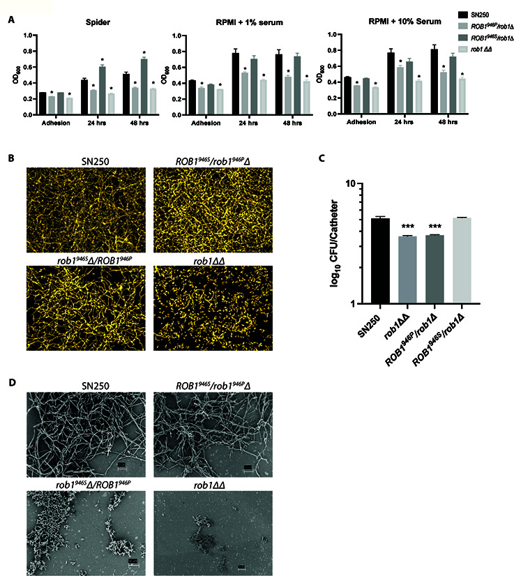
Fig 5.

The ROB1 alleles have distinct biofilm formation phenotypes in vitro and in vivo. (A) The biofilm formation of ROB1 heterozygotes was compared to SN250 and the rob1∆∆ mutant in Spider medium, RPMI + 1% serum, and RPMI + 10% serum at the indicated time points. The asterisks indicate statistically significant changes from SN250 using Student’s t test corrected for multiple comparisons (adjusted P < 0.05). (B) The biofilms were imaged using a microtiter plate imaging system as described in Materials and Methods. The apical views are shown and are representative of two replicates. (C) The fungal burden of intravascular catheters infected with the indicated strains 24 hours post infection. The bars indicate mean fungal burden from catheters placed in three rats and the error bars indicate standard deviation. The asterisk indicates statistically significant differences from WT by ANOVA followed by Dunnett’s correction for multiple comparisons (adjusted P-value < 0.05). (D) Scanning electron microscopy of biofilms formed by the indicated strains in the vascular catheters 24 hours post infection.
