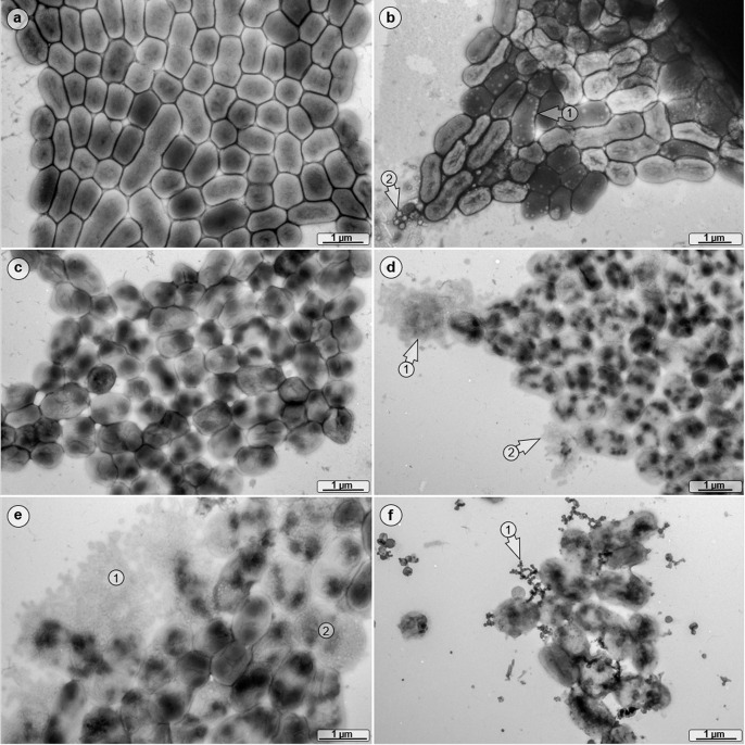
Fig 4.
Morphological analyses of VBNC cells. ATCC 19606T was grown in either low- (a) or high-salt (b) mineral medium, and TEM images were obtained at T0 and 4 days PSP. High-salt-treated cells produced numerous intracellular vesicles (arrow 1), and extracellular cell debris was detected also (arrow 2). (c) The cells of the low-salt culture 4 days PSP exhibited an altered cell morphology. (d) In the high-salt culture 4 days PSP, cytoplasm-leaking bacteria were detected (arrows 1 and 2) and (e) a high percentage of disintegrated bacterial cells was present (areas 1 and 2). (f) Resuscitated cells formed many short dendritic, globular strands as an integral part of bacterial clusters (arrow 1).

