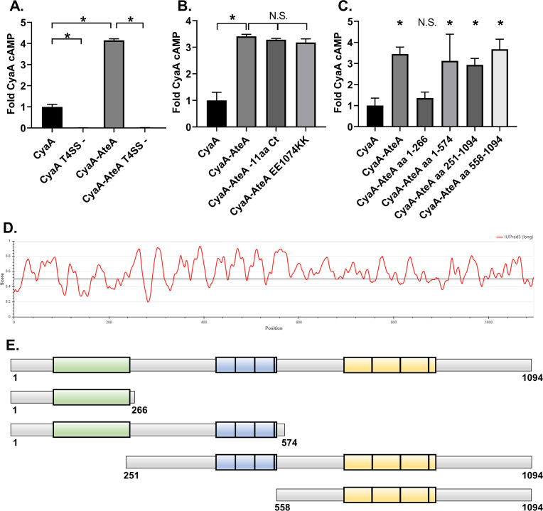
Fig 3.
AteA is recognized and secreted by a T4SS. (A–C) THP-1 cells were infected with a L. pneumophila strain expressing the indicated Cya-fusion proteins for 1 h. cAMP concentrations were quantified from infected cell lysates by ELISA and compared as fold change over CyaA alone. (A) CyaA and CyaA-AteA expressed in both wild-type L. pneumophila Lp02 or T4SS-deficient Lp03 strain (T4SS−). (B) C-terminal mutants of CyaA-AteA were expressed in Lp02 and compared to CyaA alone and CyaA-AteA. (C) Truncation constructs of CyaA-AteA diagrammed in panel E were expressed in Lp02 and compared to CyaA alone and CyaA-AteA. (D) IUPred3 order/disorder plot of AteA protein. (E) Diagram of AteA truncation mutants. Potential globular region highlighted in green, two tandem repeat regions highlighted in blue and yellow. (A–C) Error bars represent ±SD of the mean of three biological replicates with two technical replicates each, and graph is representative of two repeated experiments. *P < 0.05 (one-way ANOVA).

