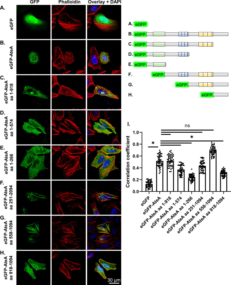
Fig 5.
Localization to actin is dependent on the second repeat region, but other regions of AteA influence the pattern. (A–H) Upper right: schematic of eGFP-AteA fusion constructs and truncations used in transfections. (A–H) Left: confocal images of HeLa cells transiently transfected to express eGFP, eGFP-AteA, or an eGFP-AteA truncation construct as diagramed. Cells were stained to visualize actin using Alexa Fluor 564 phalloidin at 36–48 h post transfection. (I) Relative co-localization with phalloidin as measured by Pearson’s correlation (n = 50 cells/group from three independent experiments). Compared by one-way ANOVA (*P < 0.05 and ns; not significant).

