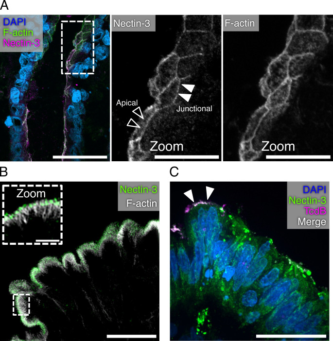
Fig 3.
NECTIN-3 localizes to the brush border of the colonic epithelium. (A) Confocal image of human colonic tissue stained for Nectin-3, F-actin, and DAPI. Dashed boxes represent the origin of the zoomed image. Black arrowheads indicate a signal localized to the apical surface of cells, while white arrowheads indicate areas of junction. Scale bars, 50 µm and 20 µm (zoom). (B) Structured illumination microscopy image of human colonic epithelium stained for Nectin-3 and F-actin. Dashed boxes represent the origin of zoomed image. The main image is presented as a max intensity projection, while the zoomed panels are shown as a single Z-plane. Scale bars, 10 µm and 2 µm (zoom). (C) Confocal image of a human colonic explant intoxicated with JF669-TcdB1 for 1 hour at 37°C and stained for Nectin-3. Scale bar, 30 µm.

