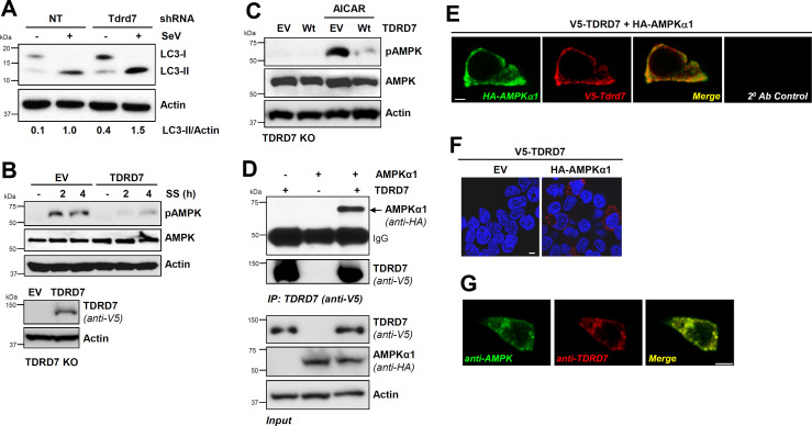
Fig 1.
TDRD7 interacts with AMPK and inhibits its activation by various stimuli. (A) L929 cells expressing Tdrd7-specific shRNA were infected with SeV, and LC3-II was analyzed by immunoblot at 8 hours post-infection (hpi). (B) TDRD7 knockout (KO) cells, ectopically expressing V5-TDRD7, were serum starved and analyzed for pAMPK by immunoblot. (C) TDRD7 KO cells, ectopically expressing V5-TDRD7, were treated with AICAR and analyzed for pAMPK by immunoblot 2 h post-treatment. (D) HEK293T cells were co-transfected with V5-TDRD7 and HA-AMPKα1 plasmids. TDRD7 was immunoprecipitated from the cell lysates 24 h post-transfection, and the immunoprecipitates were analyzed for AMPK by immunoblot. (E) HEK293T cells were co-transfected with V5-TDRD7 and HA-AMPKα1 plasmids. TDRD7 and AMPK were immuno-stained and analyzed by confocal microscopy. (F) HEK293T cells were co-transfected with V5-TDRD7 and HA-AMPKα1 plasmids, and TDRD7:AMPK interaction was analyzed by duolink assay; the red dots indicate the duolink signals. (G) HT1080 cells were immuno-stained using anti-TDRD7 and antiAMPK antibodies and analyzed by confocal microscopy. NT, nontargeting, EV, empty vector; scale bar, 5 µm. The results are representative of at least three experiments.

