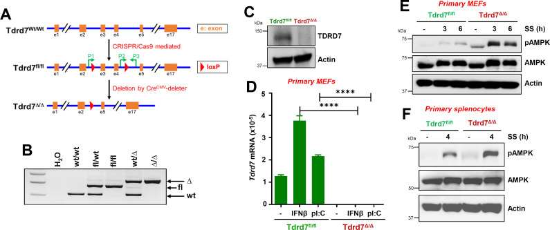
Fig 6.
Increased AMPK activation in Tdrd7 knockout primary mouse cells. (A, B) A diagram showing the newly generated Tdrd7fl/fl mice and the derived Tdrd7Δ/Δ strain (A) and the genotypes of relevant strains (B). The location of primers (P) used for genotyping is shown in A. (C) Liver homogenates from Tdrd7fl/fl and Tdrd7Δ/Δ mice were analyzed for TDRD7 by immunoblot. (D) Tdrd7fl/fl and Tdrd7Δ/Δ primary MEFs were treated with IFNβ or polyI:C for 16 h, and the Tdrd7 mRNA levels were analyzed by quantitative reverse transcription-polymerase chain reaction (qRT-PCR). (E) Tdrd7fl/fl and Tdrd7Δ/Δ primary MEFs were serum starved (SS) for the indicated times, and pAMPK was analyzed by immunoblot. (F) Tdrd7fl/fl and Tdrd7Δ/Δ primary splenocytes were serum starved (SS) and analyzed for pAMPK by immunoblot. The results are representative of three experiments using two to four mice from each group; the data represent mean ± SEM; *P < 0.05.

