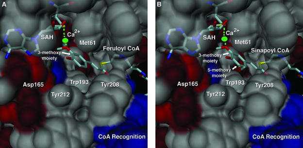
Figure 4.
Close-up molecular surface view of the CCoAOMT active site. A, The active site view includes a Ca2+ ion (green), with SAH and feruloyl CoA shown as color-coded bonds. Residues surrounding the phenylpropanoid portion of feruloyl CoA are illustrated as molecular surfaces and labeled accordingly. Surfaces are color coded according to their properties (acidic residues in red, basic residues in blue, neutral or hydrophobic residues in gray). B, The same view as in A for the sinapoyl CoA complex. Both the 3-methoxy and 5-methoxy portions of the sinapoyl product are labeled. This figure was produced with DINO (Visualizing Structural Biology, 2002, http://www.dino3d.org).

