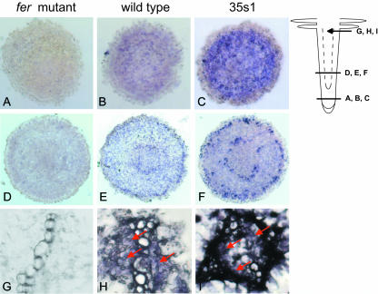
Figure 4.
FER immunolocalization using anti-N-FER antiserum on 10-μm paraffin-embedded tomato root cross-sections of fer mutant (A, D, and G), wild-type (B, E, and H), and 35s1 (C, F, and I) plants. A to C, Cross-sections from the meristematic root zone. D to F, Cross-sections from the elongation root zone as indicated on the root scheme. G to I, Magnified views of the central cylinder from cross-sections in the root hair zone. The presence of FER protein was revealed by violet staining from indirect immunolabeling with a secondary antibody coupled to alkaline phosphatase.

