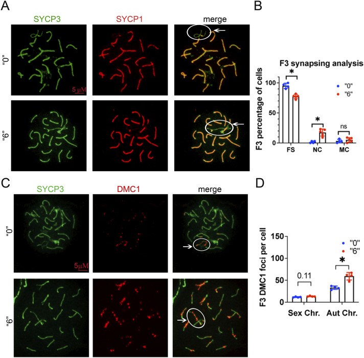
Figure 2. Meiotic defects in exposed mice.
(A) Representative image of 35-d-old F3 male testis cell spreads immunostained against central, SYCP1 (red) and lateral, SYCP3 (green) components of the synaptonemal complex, white circle indicates sex chromosomes. (B) Quantitative analysis of the synapsing defects in 35-d-old testis, FS, fully synapsed, NC, not complete, MC, multiple connections, n = 4 dose “0,” n = 4 dose “6,” non-parametric Mann-Whitney test, bar is 5 µΜ. (C) Representative image of pachytene stage immunostained by DMC1 (red) and SYCP3 (green) in control (top) and thia 6-derived (bottom) 35-d-old testis cell spreads, bar is 5 µΜ. (D) Quantitative analysis of DMC1 foci in F3 35 d-old generation males, sex and autosome (Aut.) chromosome (Chr.) foci were counted independently and compared with control samples, n = 4 dose “0,” n = 4 dose “6,” *P < 0.05, nonparametric Mann–Whitney test. All plots on the figure represent an averaged value ± SD.

