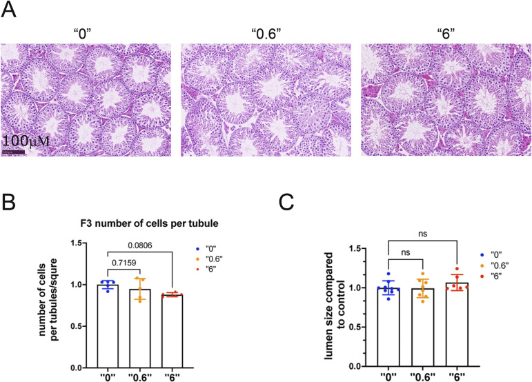
Figure S3. Testis morphology in F1 and F3 males.
Mice were euthanized at 35 d of age. The testes were fixed in Bouin’s solution and embedded in paraffin. Paraffin sections were stained with H&E and cells were counted in stage six to seven tubules. (A) Representative images of testes from mice derived from treatment doses of 0, 0.6 and 6 mg/kg/day, bar is 100 µΜ. (B) Number of cells per tubule in F3. (C) Lumen size in F3 males, F3, n = 6 dose “0,” n = 7 dose “0.6,” n = 6 dose “6,” Kruskal–Wallis test. All plots on the figure represent an averaged values ± SD.

