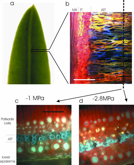
Figure 1.
A, Distal half of a 12-mm wide P. grayi leaf showing single vein. B, Fluorescence image of a paradermal section of leaf after infusion with Texas red. Tissues from left to right are leaf vein metaxylem (MX; red), transfusion tissue (TT; red), and ATT (yellow). Those ATT tracheids not containing dye appear blue due to lignin autofluorescence. C and D, Frozen cross sections of the leaf cut along the axis marked in B, showing ATT tracheids in blue (due to autofluorescence) and chlorophyll as red. Tracheids under small tensions appeared round in cross section (C) while those in water stressed leaves (D) became highly flattened. Scale bars in B = 200 μm and in C and D = 100 μm).

