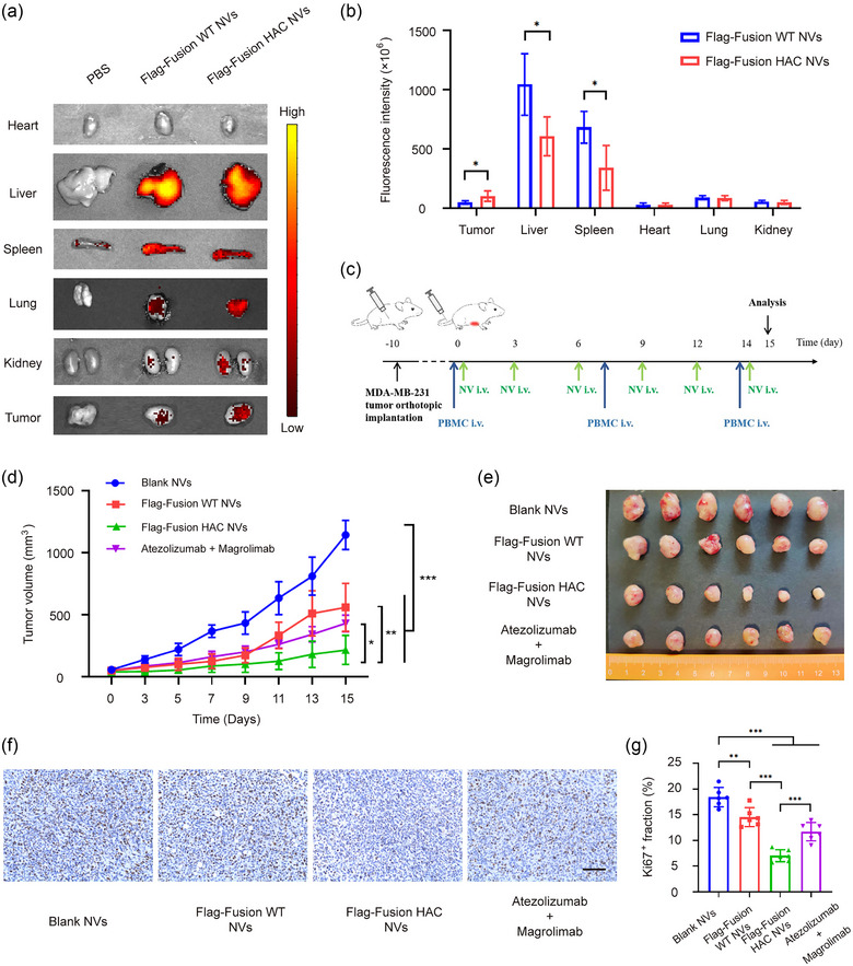
FIGURE 6.

Dual blockade of CD47 and PD‐L1 with HAC NVs inhibits tumour proliferation in vivo. (a) Representative ex vivo fluorescent images of the DiR‐labelled Blank, Flag‐Fusion WT or Flag‐Fusion HAC NVs in tumours and main organs. (b) Statistical analysis of fluorescence intensity of different organs and tumours in (a) (n = 5, *p < 0.05). (c) Schematic schedule of therapeutic study in MDA‐MB‐231 xenograft mouse model. (d) Tumour growth curves of MDA‐MB‐231 tumour‐bearing mice with treatment of NVs or antibodies (n = 6, *p < 0.05, **p < 0.01 and ***p < 0.001). (e) Photograph of tumour tissues obtained from different groups (n = 6). F. Ki‐67 levels in tumour tissues of each group presented in representative immunohistochemistry images. Scale bar: 50 μm. (g) Quantification of Ki‐67 levels in (f) (n = 6, **p < 0.01, and ***p < 0.001).
