FIGURE 7.

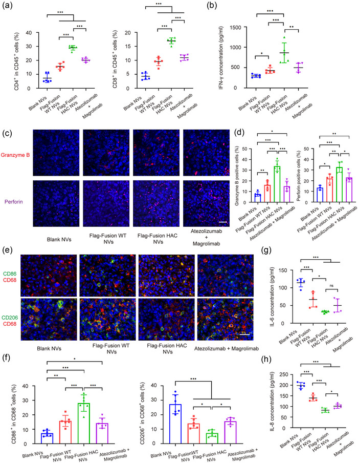
Dual blockade of CD47 and PD‐L1 with HAC NVs elicits potent antitumour immunity. (a) Flow cytometric quantification of CD4+ and CD8+ T cells in tumour from each group after indicated administration (n = 6, ***p < 0.001). (b) ELISA measurement of IFN‐γ in plasma from each group after indicated administration (n = 5, *p < 0.05, **p < 0.01 and ***p < 0.001). (c) Representative immunofluorescence images of granzyme B and perforin in tumour tissues after indicated administration (Red: granzyme B; Magenta: perforin; Blue: DAPI). Scale bar: 50 μm. (d) Statistical analysis of granzyme B and perforin in tumour samples in (c) (n = 3–5, *p < 0.05, **p < 0.01 and ***p < 0.001). (e) Representative immunofluorescence images of CD86+ M1 and CD206+ M2 macrophages in tumour tissues after indicated administration (Red: CD68; Green: CD86, CD206; Blue: DAPI). Scale bar: 50 μm. (f) Statistical analysis of M1 and M2 macrophages in tumour samples in (e) (n = 6, *p < 0.05, ***p < 0.001). (g and h) ELISA measurement of IL‐6 (g) and IL‐8 (h) in plasma from each group after indicated administration (n = 5, *p < 0.05, ***p < 0.001).
