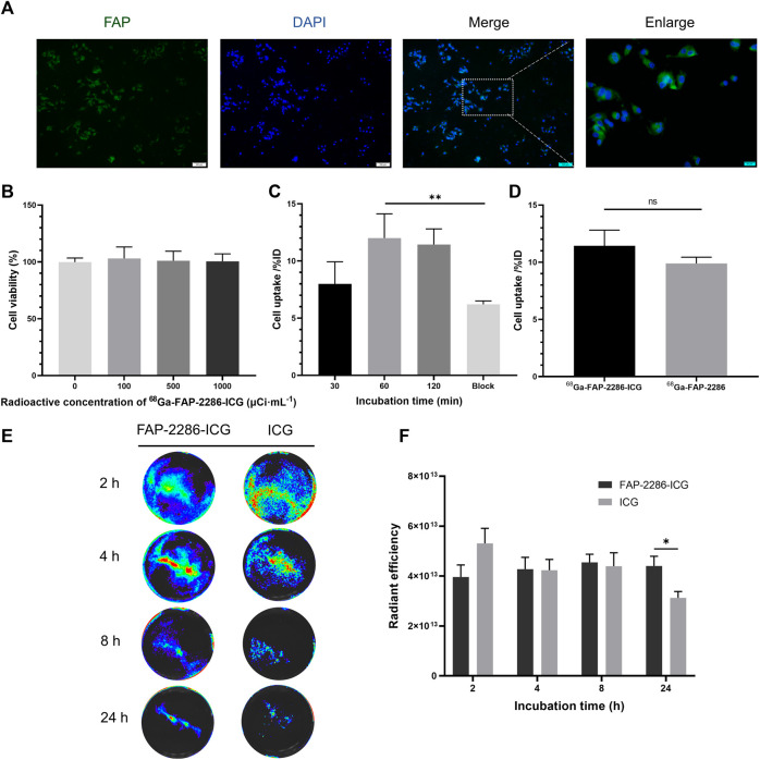
FIGURE 3.
(A) Representative immunofluorescence staining of FAP protein in U87MG cell. FAP was indicated by positive staining (green), and nuclear condensation was indicated by DAPI nuclear staining (blue). Scale bar: 100 μm; (B) Viability of U87MG cells incubated with the different radioactive concentrations of [68Ga]Ga-FAP-2286-ICG for 24 h (n = 3); (C) The radioactivity uptake of [68Ga]Ga-FAP-2286-ICG in U87MG cells; (D) Cellular uptake of [68Ga]Ga-FAP-2286-ICG compared to [68Ga]Ga-FAP-2286 in U87MG cells; Representative optical image (E) and quantification (F) of U87MG cells interaction after incubation for 2, 4, 8, and 24 h with 3 μM of either FAP-2286-ICG or ICG (n = 5). ns: not statistically significant; *p < 0.05; **p < 0.01.

