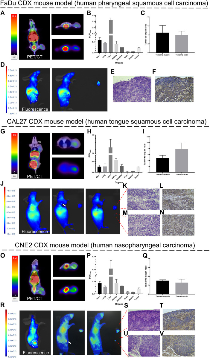
FIGURE 6.
Proof-of-concept of diagnosis and fluorescence-guided surgery studies in HNCs tumor-bearing mice. Representative PET/CT images (1 h post-injection) and fluorescence images (72 h post-injection) of [68Ga]Ga-FAP-2286-ICG in FaDu (A, D), CAL27 (G, J), and CNE2 (O, R) tumor-bearing mice. White arrows indicate residual fluorescence signal in the surgery bed; SUVmax of critical organs in FaDu (B), CAL27 (H) and CNE2 (P) tumor-bearing mice at 1 h post-injection; Tumor-to-organ ratios of [68Ga]Ga-FAP-2286-ICG in FaDu (C), CAL27 (I) and CNE2 (Q) tumor-bearing mice at 1 h post-injection; pathology and FAP immunohistochemistry of resected tissue of FaDu (E. F), CAL27 (K–N) and CNE2 (S–V) tumor-bearing mice confirmed the presence and positive staining of FAP in tumor stroma (stained brownish yellow); magnification × 200. Scale bar: 50 μm. Data were expressed as mean ± SD (n = 3).

