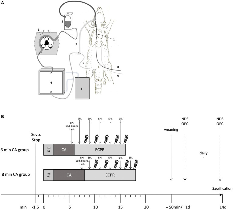
Figure 1.
(A,B) ECMO set-up in the rat and Timeline of the experimental protocol. (A) Extracorporeal membrane oxygenation (ECMO) circuit consisting of a venous ECMO drainage cannula (1) inserted in the right jugular vein, an open reservoir (2), a roller pump (3), an oxygenator (4) with attached heat exchanger (5) and an arterial ECMO cannula (6) inserted in the right femoral artery. All these elements (custom-made; Dipl.-Ing. Martin Humbs, Valley Germany) were connected by a tubing system with shortcut (7) for priming before resuscitation. Medication was given via left femoral venous cannula (8), invasive blood pressure was measured and blood gas samples collected via left femoral arterial cannula (9). (B) Timeline of the experimental protocol in the 6 min and 8 min cardiac arrest (CA) group; Sevoflurane stop (Sevo. Stop) 1.5 min bevor inducing of ventricular fibrillation (ind VF); Followed by cardiac arrest (CA) for 6 or 8 min; Extracorporeal cardiopulmonary resuscitation (ECPR); heparin (Hep.); epinephrine (EPI.); sodium bicarbonate (Sod. bicarb.); daily scoring of Neurologic Damage Score (NDS) and Overall Performance Category (OPC); sacrification at day 14.

