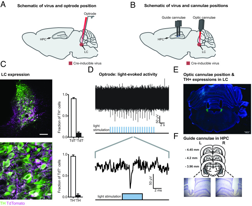
Fig. 2.
Histological confirmation and optogenetic activation of LC-TH+ neurons. (A) Schematic showing the placement of an optrode (optic fiber and electrode) used to record light-evoked multiunit firing in LC-TH+ neurons of Th-Cre rat expressing Cre-inducible AAV-DJ ChrimsonR-tdTomato (ChR+) virus. (B) Schematic showing the placement of bilateral optic cannulae in the LC and bilateral guide cannulae in the dorsal hippocampus (HPC) of Th-Cre rat. (C) Overlap between TH+ neurons and neurons expressing ChrimsonR-tdTomato at the position of the optic cannula in the LC. (Scale bars: Upper, 100 μm; Lower, 10 μm.) (D) Example of multiunit responses to optical stimulation recorded in the LC. The Upper panel shows multiunit spikes elicited by a train of light stimuli (20 5-ms pulses delivered at 1 Hz; note that each light pulse elicits a response). The Lower panel shows an expanded view of the multiunit response to a single pulse of light stimulation. (E) A representative example of the bilateral optic cannula positions in the LC used in behavioral experiments. (Scale bar: 1 mm.) (F) Histological verification of the bilateral guide cannulae tip locations in HPC used for drug infusions.

