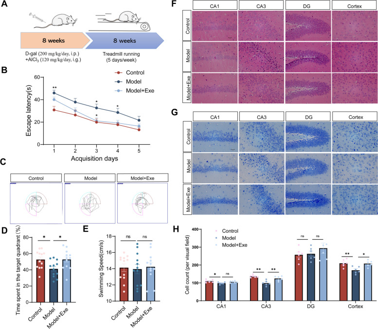
Fig. 1.
Exercise improves cognitive function and reverses hippocampal neuron loss in AlCl3/D-gal-treated mice. A Flow chart for the experimental design. B The escape latency of mice to the platform during the acquisition phase (n = 12 per group). C Representative track images of each group mice in day 6 probe trial test. D Time spent in the target quadrant (%) within 60 s of mice in the probe trial (n = 12 per group). E The mean swimming speed of each group mice in day 6 probe trial test (n = 12 per group). F Representative images of HE staining in various brain regions (magnification 400 ×). G Representative images of Nissl staining in various brain regions (magnification 400 ×). H The number of living neurons of hippocampus and prefrontal cortex in mice (n = 6 per group). Data are means ± SEM. *p < 0.05, **p < 0.01 vs. Control group; *p < 0.05 in vs. Model group in B. *p < 0.05, **p < 0.01, ns-not significant in D, E and H. Statistical analysis was performed using two-way (B) and or one-way (D, E, H) ANOVA, followed by Tukey’s multiple comparisons test

