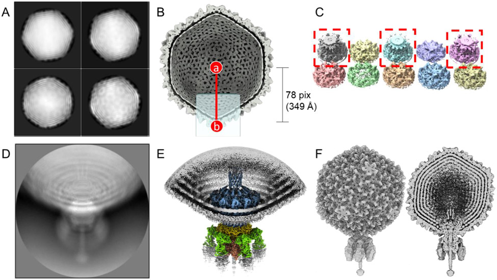
Figure 1. The pipeline of localized reconstruction.
A. Representative 2D class averages of the S6 virion calculated using particles binned 4 times. B. Section view of the S6 icosahedral reconstruction with the 5-fold icosahedral axis aligned to the Z-axis (I3/I4 convention). Position (a) is the center of the icosahedral capsid, while (b) is the center of the re-extracted particle, re-centered from the icosahedral head. The light-blue square represents the position of the cylindrical mask, which covers the potential tail location. C. Ten 3D classes were obtained by non-sampling 3D classification of icosahedral expanded particles. 3D classes containing the Sf6-aligned tail are boxed in red. D. A representative 2D class average obtained from re-extracted tail particles. E. Localized reconstruction of aligned tail particles after applying C6 symmetry. F. Asymmetric reconstruction of the entire Sf6 virion (left panel) with both tail- and capsid-aligned particles. The right panel shows a section through the virion.

