Fig. 1.

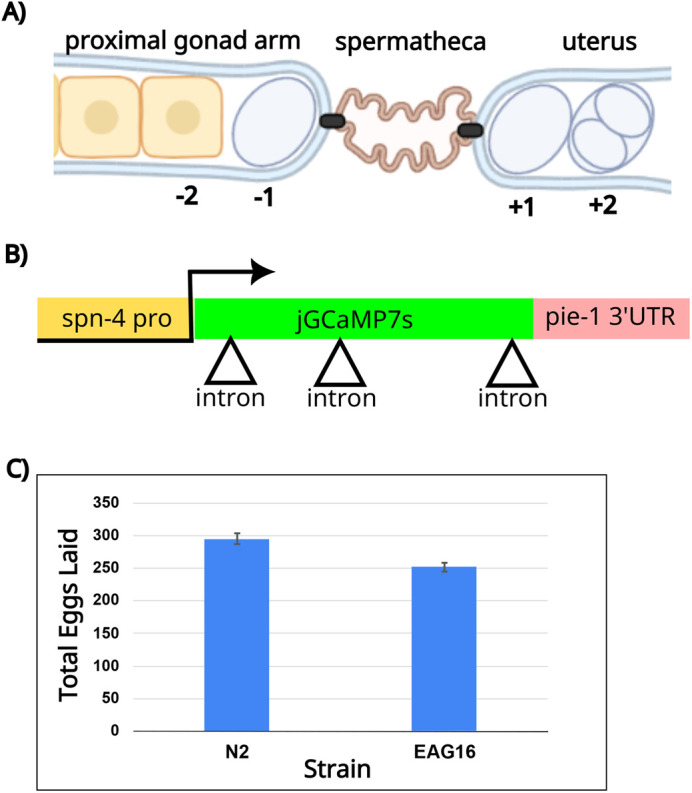
Design strategy for a fertilization-specific GECI. (A) Schematic of the worm gonad featuring a portion of the proximal gonad arm with a single-file row of developing oocytes, the spermatheca where the sperm are stored, and the uterus where embryos are stored temporarily before egg lay. The proximal oocyte has undergone meiotic maturation (shown by a change in shape and nuclear envelope breakdown) and is designated −1. The newly fertilized embryo in the uterus is labeled +1. Image created using Biorender.com. (B) Schematic of reporter design showing the spn-4 promoter, the jGCaMP7s coding sequence interrupted by three introns (triangles) and the pie-1 3′ UTR. (C) Brood size analysis of the EAG16 reporter strain compared to N2 wildtype worms. Error bars indicate the s.e.m.
