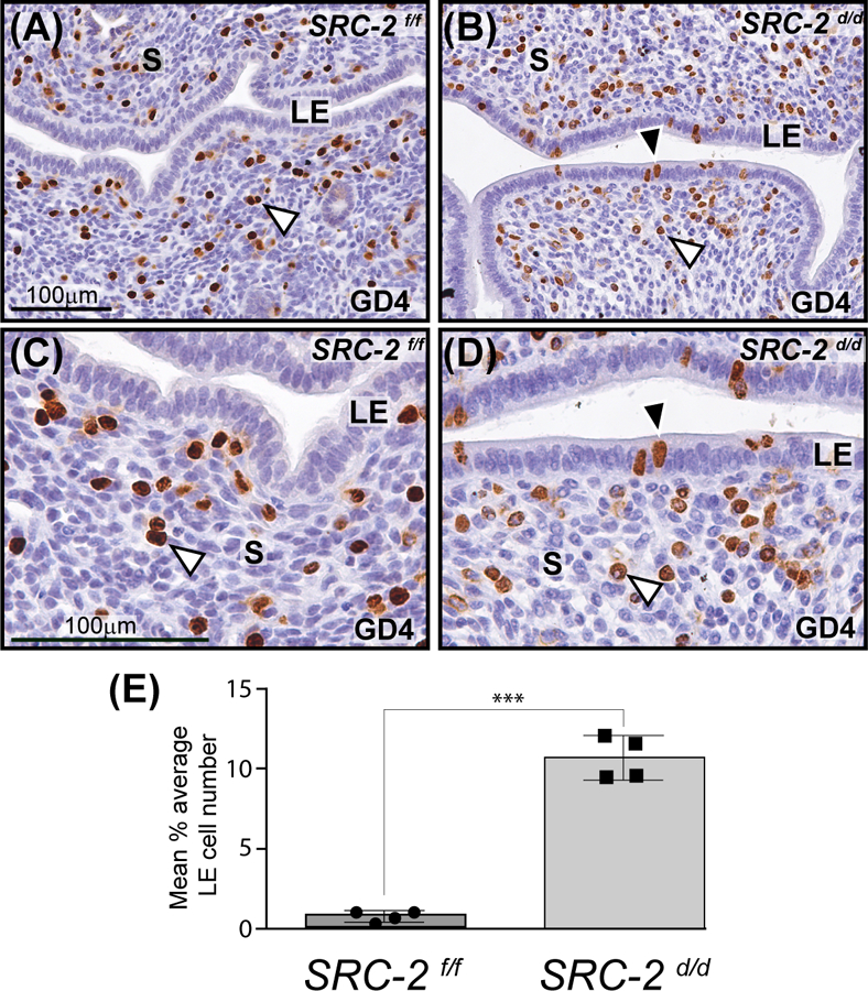
FIGURE 2.

Retention of a subset of proliferating luminal epithelial cells in the SRC-2 d/d endometrium at GD4. (A) Immunohistochemical detection of cells that are positive for BrdU incorporation in the endometrium of the SRC-2 f/f at GD4. Note the presence of proliferating stromal cells in the endometrium of the SRC-2 f/f mouse at GD4 (white arrowhead). In contrast, the luminal epithelium (LE) of the SRC-2 f/f endometrium is negative for BrdU immunopositivity. Together, the epithelial and stromal cellular proliferative profiles are indicative of a receptive uterus at GD4 47. (B) A subset of luminal epithelial cells in the SRC-2 d/d endometrium at GD4 is consistently immunopositive for BrdU incorporation (black arrowhead); a subgroup of subluminal stromal cells is positive for BrdU incorporation (white arrowhead). Panels (C) and (D) are higher power magnifications of regions shown in panels (A) and (B) respectively. Again note the absence and presence of BrdU immunopositive luminal epithelial cells in the SRC-2 f/f and SRC-2 d/d endometrium respectively. Although qualitative, BrdU positive stromal cells in the SRC-2 f/f endometrium consistently register a stronger immunopositive signal than BrdU positive stromal cells in the SRC-2 d/d endometrium, compare the stromal (S) compartment in (C) with (D). Scale bar in (A) and (C) apply to (B) and (D) respectively. (E) Histogram graphically displays the average number of BrdU positive cells per 100 luminal epithelial cells counted from each of three separate tissue sections per mouse (4 mice per genotype).
