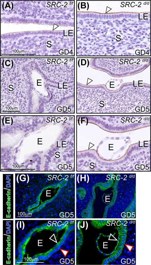
FIGURE 3.

Luminal epithelial (LE) cell marker expression is altered in the SRC-2 d/d endometrium. (A) An endometrial tissue section obtained from a SRC-2 f/f mouse at GD4; section is immunohistochemically stained for MUC 1 expression. Note the low levels of MUC 1 expression on the apical surface of the LE (white arrowhead). (B) Expression of MUC 1 is noticeably stronger on the apical surface of the LE of the SRC-2 d/d endometrium at GD4 (white arrowhead). (C) By GD5, MUC 1 expression is absent in the endometrium of the SRC-2 f/f mouse; E indicates embryo. (D) At GD5, the SRC-2 d/d endometrium still retains strong MUC 1 immunopositivity on the apical surface of the LE (white arrowhead) despite the presence of an embryo. (E) and (F) show higher power magnification images shown in (C) and (D) respectively. Again note the absence of MUC 1 expression in the LE of the SRC-2 f/f endometrium at GD5 and the continued presence of MUC 1 expression on the apical surface of the LE compartment within the SRC-2 d/d endometrium (white arrowhead). (G) Immunofluorescence detection of E-cadherin in the epithelium of the SRC-2 f/f endometrium at GD5; E denotes embryo. Note E-cadherin expression is specific to the epithelial chamber of the SRC-2 f/f implantation site. (I) Higher power magnification image of region shown in (G). Note that E-cadherin expression is present in the trophectoderm of the embryo (black arrowhead) whereas there is significantly less E-cadherin expression in the basolateral regions of epithelial cells that are juxtaposed to the embryo (white arrowhead). (I) Immunofluorescence detection of the E-cadherin protein in the SRC-2 d/d endometrium at GD5. As in (G), E-cadherin immunopositivity is specifically localized to the epithelial compartment. (J) Higher power magnification image of a region shown in (H). Note the retention of E-cadherin expression in the apical and basolateral regions of the epithelial cells (white arrowhead); trophoblast cells of the embryo are positive for E-cadherin expression (black arrowhead). Scale bar in (G) and (I) apply to (H) and (J) respectively.
