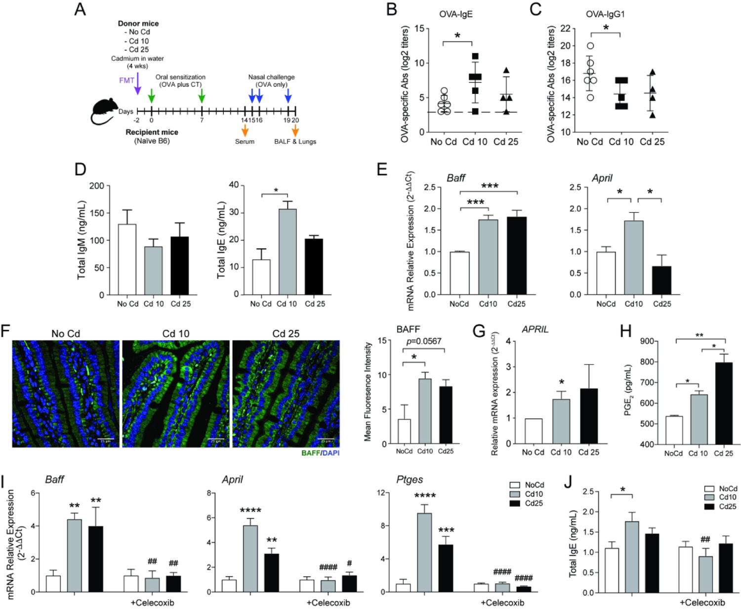
Figure 5. Cd-induced gut microenvironment enhances IgE responses via stimulation of PGE2.

(A) Experimental scheme for the transfer of fecal materials from Cd-treated mice. Allergen-specific serum IgE (B) and IgG1 (C) responses after oral sensitization of recipient mice not exposed to Cd. (D) IgM and IgE production in culture supernatants of naïve murine spleen cells cultured for 4 days in the presence of IL-4 (10 ng/ml), anti-CD40 (1μg/ml), and bacteria-free fecal extracts (n = 4). (E) Baff and April mRNA responses in Myd88 KO macrophages cultured 24 h in the presence of bacteria-free fecal extracts (n=5). (F) Expression of BAFF in small intestinal tissues of naïve mice or mice exposed to subtoxic doses of Cd for 28 days (n=5). (G) PGE2 secretion from HT-29 cells 4 h after the addition of bacteria-free fecal extracts (n=5). (H) Baff, April, and Ptges mRNA responses by MLN lymphocytes (2×10⁶ cells/mL) stimulated for 24 h with anti-CD40 and IL-4 in the presence of bacteria-free fecal extracts only, or together with the COX2 inhibitor celecoxib (10 μM) (n=3). (I) Effects of celecoxib on IgE production by MLN lymphocytes stimulated as described in (H) (n=3). Data are from at least four independent experiments and are expressed as the mean ± SD. *p < 0.05; **p < 0.01; ***p < 0.001; ****p < 0.0001 compared to NoCd. ## p < 0.01; ##p < 0.001; ####p < 0.0001 compared to no celecoxib.
