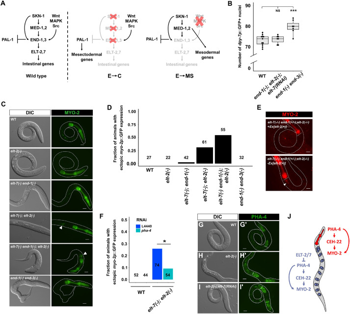
Fig. 4.
ELT-2 and ELT-7 repress pharyngeal gene expression in the intestine. (A) E→C or E→MS binary fate choice during early embryogenesis. PAL-1 is required for the specification of the C blastomere, which gives rise to epidermis and body wall muscles (Hunter and Kenyon, 1996). Maternally provided pal-1 is specifically translated in EMS and P2. In MS and E, PAL-1 activity is blocked by TBX-35 and END-1/3, respectively (Broitman-Maduro et al., 2006). Thus, depleting TBX-35, END-1/3 or their upstream activators, MED-1/2 and SKN-1, causes excess skin and muscle owing to the misspecification of MS and/or E into C, the somatic descendant of P2 (Hunter and Kenyon, 1996). As EMS divides, Wnt/MAPK/Src signaling from P2 polarizes EMS, leading to the phosphorylation of POP-1 and activation of end-1/3 in E, but not in MS. When the polarizing signal from P2 is disrupted, POP-1 is unphosphorylated and MED-1/2 instead activate the development of MS, which gives rise to the posterior pharynx and body wall muscles (Maduro and Rothman, 2002; Maduro et al., 2002; Rocheleau et al., 1999; Shin et al., 1999). (B) Both WT and end-1(−);elt-2(−);elt-7(RNAi) larvae contain ∼73 epidermal cells, whereas end-1(−) end-3(−) larvae contain ∼80 epidermal cells marked by dpy-7p::GFP. Boxes represent the 25-75th percentiles and the median is indicated. The whiskers show the 1.5× interquartile range. NS, not significant (P>0.05); ***P≤0.001 (parametric one-way ANOVA followed by pairwise two-tailed unpaired t-tests with Benjamini–Hochberg correction). (C) Representative differential interference contrast (DIC) and fluorescence micrographs showing different mutation combinations expressing myo-2p::GFP. Arrowheads indicate ectopic expression of myo-2 in elt-7(−);elt-2(−) and elt-7(−) end-1(−);elt-2(−). (D) The frequency of animals showing mis-expression of myo-2, as shown in C. The number of animals scored for each genotype is indicated. (E) Fluorescence micrographs showing expression of myo-2p::mCherry present on tmC12, which balances end-1(−). An elt-7(−/−) end-1(+/−);elt-2(−/−) animal that has lost the elt-2(+) rescuing array shows ectopic expression of myo-2 in the midgut (arrowhead) (n=50). The white horizontal lines mark the posterior end of the pharynx. (F) Knocking down pha-4 partially rescues ectopic expression of myo-2 in elt-7(−);elt-2(−) animals. *P≤0.05 (Fisher's exact test). (G-I) The expression of the endogenously tagged pha-4 reporter in WT (G,G′) (n=120), elt-2(−) (H,H′) (n=40) and elt-2(−);elt-7(RNAi) (I,I′) (n=30) animals. The white horizontal lines in G′-I′ mark the posterior end of the pharynx. Exposure time: 195 ms. All scale bars: 10 μm. (J) Model of spatial repression and fate exclusion in the digestive tract.

