Fig. 3.
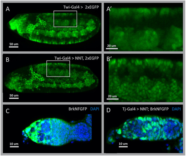
NaNuTrap efficiently transfers zygotically expressed cytoplasmic FP into the nucleus in vivo. (A) TTG (Twi-Gal4, UAS-2×EGFP) balancer line embryo at stage 11. A large amount of EGFP is expressed in mesoderm derivative cells. (A′) Higher magnification of the area indicated in A. (B) Additional to cytoplasmic EGFP, NNT is also expressed by the Twi-Gal4 driver of the TTG (TM3, Twi-Gal4, UAS-2×EGFP) construct in the embryo shown at stage 11. (B′) Higher magnification of the area indicated in B. (C) Fixed tissue sample of germarium, anterior to the left, showing GFP expression driven by the large reporter construct BrkNFGFP (green; Dunipace et al., 2013) and nuclei stained with DAPI (blue). (D) The BrkNFGFP reporter combined with NNT and a pan follicle cell driver, Tj-Gal4, show distinct nuclear localization of GFP within cells.

