Fig. 6.
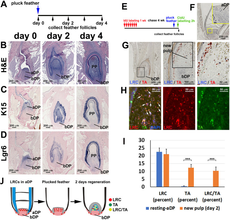
Long-term label-retaining dermal cells in the apical dermal papilla contribute to pulp regeneration. (A) Strategy to collect regenerating feather follicles at different time points. (B) H&E staining of feather follicles. (C,D) K15 (C) and Lgr6 (D) in situ hybridization. (B-D) Left column, feather follicles after plucking at resting phase; middle column, 2 days after plucking; right column, 4 days after plucking. (E) Strategy of LRDCs and TA cell double labeling with IdU and CldU, respectively. (F) IdU staining of a resting-phase feather follicle showing the LRCs in the aDP. (G) Double staining of LRDCs (blue) and TA cells (red) in a follicle regenerating for 2 days. (H) Fluorescent immunostaining of LRDCs (red) and TA cells (green). White arrows indicate the double-labeled new PP cells. (I) Percentage of CldU-positive cells (TA cells), IdU-positive cells (LRCs), and double labeled CldU/IdU-positive cells in the resting-phase aDP and new pulp after 2 days of regeneration (n=4 feather follicles, ***P<0.001, paired two-tailed Student's t-test). Data are mean±s.d. (J) Schematic showing LRDCs in aDP participating in pulp regeneration. aDP, apical dermal papilla; bDP, basal dermal papilla; PP, pulp.

