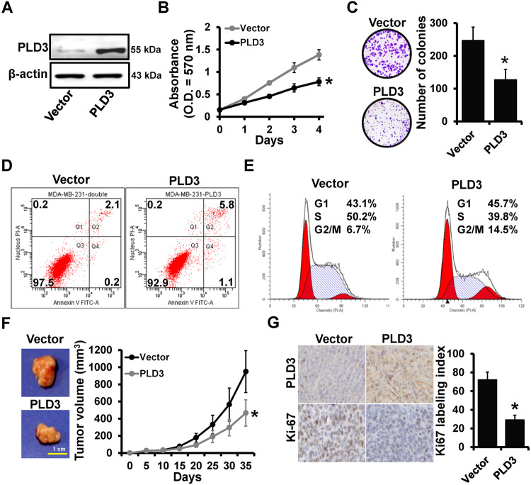
Fig. 1. PLD3 functioned as a tumor suppressor gene in breast cancer.
A PLD3 expression in stable PLD3-overexpression cells (231-PLD3) and control cells (231-Vector) was determined by the western blot. MTT (B) and colony-formation (C) analyzed the proliferation of the cells described in (A). Flow cytometry analyzed the apoptosis (D) and the cell cycle distribution (E) of the cells described in (A). F Tumor growth curves of the subcutaneous tumor made of the cells described in (A) at the indicated times and dissected tumors photographed at the harvest time, each group included 6 mice. G IHC staining analyzed the expression level of Ki-67 in breast cancer tissues with high or low PLD3 expression, the representative IHC image of each mouse has been selected for statistical analysis. *P < 0.05.

