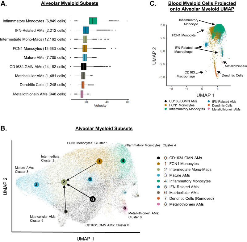
Fig. 3. Intermediate monocyte-macrophage subsets are present in the lung.
A Box-plots of median (center line), interquartile range (edge of box), 1.5x interquartile range (whiskers), and individual outliers (dots) of RNA velocity for each alveolar myeloid subset. B Partition-based graph abstraction (PAGA) of RNA velocity field projected on the alveolar myeloid UMAP (Fig. 1B). Gray dotted lines represent topologic connectivity of subsets. Arrows represent RNA velocity trajectory-inference (alveolar macrophage = AM). Dendric cells were excluded from RNA velocity analysis. C We collected paired peripheral blood mononuclear cells (PBMC) from participants who underwent research bronchoalveolar lavage (BAL). We isolated single cells and assessed them with CITE-Seq. We selected cells that mapped to blood myeloid lineage markers (monocytes, macrophages, and DCs) and then projected them into the BAL UMAP space. Blood monocytes clustered in the upper right of the BAL UMAP (occupying the same BAL UMAP space as FCN1 Alveolar Monocytes and Inflammatory Alveolar Monocytes). Blood DCs occupied the same BAL UMAP space as alveolar DCs.

